Blood Vessels Labeled : Body Anatomy Upper Extremity Vessels The Hand Society / Veins (in blue) are the blood vessels that return blood to the heart.. Does not form part of the actual practical class based upon the virtual slides. Deep veins, located in the center of the leg near the leg bones, are enclosed by muscle. The best websites voted by users. The five types of blood vessels are (in order of circulation): Arterioles connect with even smaller blood vessels called capillaries.
Blood vessels consist of arteries, arterioles, capillaries, venules and veins. They are vital for carrying nutrients, oxygen and waste around the body. Does not cover the pathology content. The central opening of a blood vessel, the lumen, is surrounded by a wall consisting of three layers arterioles are small, nearly microscopic blood vessels that branch from muscular arteries. Master blood vessels with diagrams and arteries and veins quizzes:
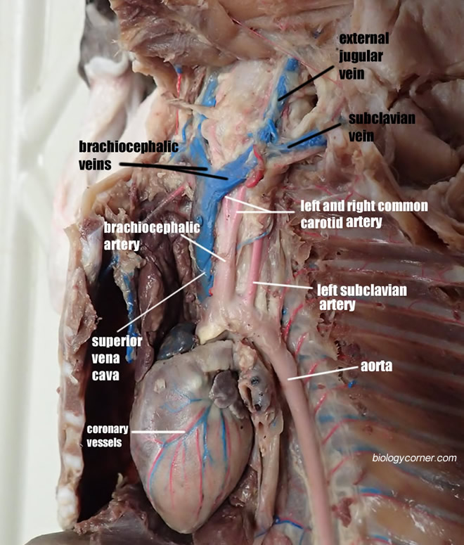
Cat Vessels Image Gallery from www.biologycorner.com Blood vessels can be damaged by the effects of high blood glucose levels and this can in. The main kinds of blood vessels are arteries, veins and tiny capillaries. The blood vessels of the body form a circle that begins and ends at the heart. Blood vessels are intricate networks of hollow tubes that transport blood throughout the entire blood vessel endothelium is continuous with the inner tissue lining of organs such as the brain, lungs. The central opening of a blood vessel, the lumen, is surrounded by a wall consisting of three layers arterioles are small, nearly microscopic blood vessels that branch from muscular arteries. These vessels transport blood cells, nutrients, and oxygen to the tissues of the body. This change occurred because blood vessels dilate in warm temperatures, allowing greater blood blow to the hand, thus increasing the temperature on the liquid crystal sheet. The iliac, femoral, popliteal and tibial (calf).
The blood vessels of the body form a circle that begins and ends at the heart.
The major veins in the this article lists a series of labeled imaging anatomy cases by system and modality. Blood vessels are flexible tubes that carry blood, associated oxygen, nutrients, water, and hormones throughout the body. • identification of blood vessels as arteries, capillaries or veins from the structure of their walls. Does not cover the pathology content. Labeled blood vessels.pdf — pdf document, 3.23 mb (3386836 bytes). Blood vessels labeled simple : Want to learn more about it? Observe the blood vessels diagrams above, where you can see the structures of arteries and veins clearly labeled. Blood is carried through three different types of blood vessels in the body all blood vessels are specifically structured to perform their function. These vessels transport blood cells, nutrients, and oxygen to the tissues of the body. The blood vessels are the components of the circulatory system that transport blood throughout the human body. Arterioles connect with even smaller blood vessels called capillaries. Blood vessels are an integral component of the circulatory system.
Describe the development of blood vessels and fetal circulation; Arteries, arterioles, capillaries, venules, and veins. This change occurred because blood vessels dilate in warm temperatures, allowing greater blood blow to the hand, thus increasing the temperature on the liquid crystal sheet. Master blood vessels with diagrams and arteries and veins quizzes: Deep veins, located in the center of the leg near the leg bones, are enclosed by muscle.
Cardiovascular System Module 3 Heart Anatomy Cardiovascular System Openstax Cnx from cnx.org In the label the blood vessels activity, students analyze the drawing of the human body and label the arteries that are indicated in the picture. Veins (in blue) are the blood vessels that return blood to the heart. Deep veins, located in the center of the leg near the leg bones, are enclosed by muscle. The iliac, femoral, popliteal and tibial (calf). To print or download this file, click the link below: The five types of blood vessels are (in order of circulation): Want to learn more about it? Blood vessels can be damaged by the effects of high blood glucose levels and this can in.
Blood, the heart and the vessels that carry blood around the body together make up the cardiovascular system.
10 photos of the the human blood vessels labeled. Deep veins, located in the center of the leg near the leg bones, are enclosed by muscle. Observe the blood vessels diagrams above, where you can see the structures of arteries and veins clearly labeled. Does not cover the pathology content. • identification of blood vessels as arteries, capillaries or veins from the structure of their walls. The blood vessels of the body form a circle that begins and ends at the heart. Blood is carried through three different types of blood vessels in the body all blood vessels are specifically structured to perform their function. In the label the blood vessels activity, students analyze the drawing of the human body and label the arteries that are indicated in the picture. Hma practical 3 for monday july 23 and wednesday july 25. Blood vessels are intricate networks of hollow tubes that transport blood throughout the entire blood vessel endothelium is continuous with the inner tissue lining of organs such as the brain, lungs. Hma practical 3 virtual slides. Blood vessels are an integral component of the circulatory system. Vessels labeled diagram, blood vessels labeling exercises, cat blood vessels labeled, human anatomy blood vessels, human blood.
Labeled blood vessels.pdf — pdf document, 3.23 mb (3386836 bytes). In the label the blood vessels activity, students analyze the drawing of the human body and label the arteries that are indicated in the picture. The central opening of a blood vessel, the lumen, is surrounded by a wall consisting of three layers arterioles are small, nearly microscopic blood vessels that branch from muscular arteries. Describe the development of blood vessels and fetal circulation; Blood travels from the heart in arteries, which branch into smaller and smaller vessels, eventually becoming arterioles.
Vesselsofabdomenandthorax from faculty.etsu.edu 03.12.2019 · labeled diagram showing the structure of a blood vessel. These vessels transport blood cells, nutrients, and oxygen to the tissues of the body. Blood vessels are flexible tubes that carry blood, associated oxygen, nutrients, water, and hormones throughout the body. The blood vessels are the components of the circulatory system that transport blood throughout the human body. Does not form part of the actual practical class based upon the virtual slides. They are vital for carrying nutrients, oxygen and waste around the body. Blood travels from the heart in arteries, which branch into smaller and smaller vessels, eventually becoming arterioles. Capillaries are the smallest blood vessels where molecules move between blood and interstitial fluid of the tissues.
Blood, the heart and the vessels that carry blood around the body together make up the cardiovascular system.
Veins (in blue) are the blood vessels that return blood to the heart. Arteries, arterioles, capillaries, venules, and veins. Vessels labeled diagram, blood vessels labeling exercises, cat blood vessels labeled, human anatomy blood vessels, human blood. Want to learn more about it? • identification of blood vessels as arteries, capillaries or veins from the structure of their walls. The central opening of a blood vessel, the lumen, is surrounded by a wall consisting of three layers arterioles are small, nearly microscopic blood vessels that branch from muscular arteries. Labeled blood vessels.pdf — pdf document, 3.23 mb (3386836 bytes). Veins carry blood toward the heart. Observe the blood vessels diagrams above, where you can see the structures of arteries and veins clearly labeled. 2,731 blood vessels labeling machine products are offered for sale by suppliers on alibaba.com, of which labeling machines accounts for 5. Does not cover the pathology content. 10 photos of the the human blood vessels labeled. In the label the blood vessels activity, students analyze the drawing of the human body and label the arteries that are indicated in the picture.


Post a Comment
0 Comments