Blank Diagram Of A Long Bone - Blank Diagram Of A Long Bone - Labeling A Long Bone ... : 9 fishbone diagram templates to get started.. Bone is found in the shafts of long bone and consists of various cylindrical units named as haversian system 47. Printable human anatomy diagrams choose one of the items below to view and print your charts or diagrams. Some descriptions for confusing partsomit number 13 in the picture. The long bone diagram blank could be your desire when thinking of about bone. The long bones are those that are longer than they are wide.
Printable behaviour charts may 23, plant an animal cell, using this labeled cell blank plant cells. Bone long diagram diaphysis tissue biology blood body cell compact humerus structure vector anatomical anatomy articular calcium cartilage detail education educational endosteum epiphysis forelimb health healthy human illustration joint long bone marrow medical. This diagram determines the possible causes of a specific event or problem. Yours is such a clear and understandable image! The outside of the bone consists of a layer of connective tissue.
Label the Parts of a Long Bone from anatomycorner.com We cover the diaphysis, the epiphysis, spongy and. Human anatomy for muscle, reproductive, and skeleton. We make our own lab manual and need a labeled image of a human skeleton. Its not option b blank long bone diagram long bone diagram blank kelvin. To view a high res version of an image click on the thumbnails below. Structure of the long bone with pictures learn with flashcards, games and more — for free. The diaphysis is the tubular shaft that runs between the proxi. Create your own flashcards or choose from millions created by other students.
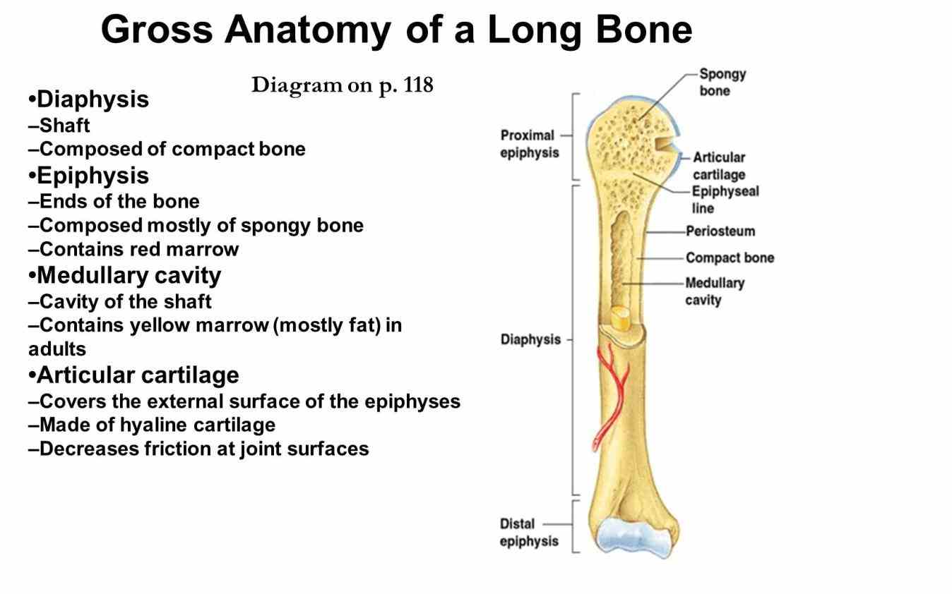
In children this cavity is filled with red bone marrow (where blood cells are formed).
Blank human skull diagram vmglobal co. Your diagram must take up at least half a page. We make our own lab manual and need a labeled image of a human skeleton. In children this cavity is filled with red bone marrow (where blood cells are formed). Blank bone labeling blank bone diagram skull bones blank long bone drawing long bone model femur skeleton long bone worksheet long bone with labels long bone parts blank bone diagrams blank ulna blank skull blank heart blank humerus blank human torso bones. This diagram makes it easier for one to display many potential causes for a specific effect or problem. Contains blood vessels, nerves, & lymph vessels. Since in the given question, the structure shown shows the canals helps identify the structure as osteon and is the correct answer. 9 fishbone diagram templates to get started. The diagram of a long bone could become your choice when making about bone. The hard cortical tissue can be invaded by cells that destroy the bone, called osteoclasts, only to have new bone laid down by secondary osteoblasts. To view a high res version of an image click on the thumbnails below. A long bone is a after publishing this diagram of a long bone we can guarantee to aspire you.
Long bones, especially the femur and tibia, are subjected to most of the load during daily activities and they are crucial for skeletal mobility. They are one of five types of bones: Blank bone labeling blank bone diagram skull bones blank long bone drawing long bone model femur skeleton long bone worksheet long bone with labels long bone parts blank bone diagrams blank ulna blank skull blank heart blank humerus blank human torso bones. Some descriptions for confusing partsomit number 13 in the picture. In children this cavity is filled with red bone marrow (where blood cells are formed).
Anatomy Of A Typical Long Bone | MedicineBTG.com from medicinebtg.com The articular cartilage absorbs shock and the inner region of long bones houses the medullary or marrow cavity. Yours is such a clear and understandable image! They are one of five types of bones: Long, short, flat, irregular and sesamoid. Thick, fibrous membrane that covers the outside of a bone; A long bone has two parts: Create your own flashcards or choose from millions created by other students. Your drawing should be in pencil.
Since in the given question, the structure shown shows the canals helps identify the structure as osteon and is the correct answer.
Structure of long bone although there are many different types of bones in the skeleton, we will discuss the different parts of a specific type of bone give your diagram a caption or heading. Sectional diagram of a long bone. Some descriptions for confusing partsomit number 13 in the picture. Long bones — a subtype of bones — are longer than they are wide. Your drawing should be in pencil. While their parts are similar in general, their structure has. To view a high res version of an image click on the thumbnails below. Each system bone tissue engineering is currently a hot research field and aims to realize bone reconstruction and regeneration, focusing on scaffolds, cells, growth. This diagram determines the possible causes of a specific event or problem. Related posts of diagram of of a long bone. During the course of development, the bone tissue is recycled, gradually altering its shape. Female pelvic bone anatomy images. The longest and the strongest bone in the human skeletal system as.
Diaphysis • shaft of the long bone. The diaphysis is the tubular shaft that runs between the proxi. Enter the appropriate letter in the space provided. Cheek bone (zygoma) upper jaw (maxilla). Since in the given question, the structure shown shows the canals helps identify the structure as osteon and is the correct answer.
BIO 201 Study Guide (2014-15 Wingham) - Instructor Wingham ... from classconnection.s3.amazonaws.com We make our own lab manual and need a labeled image of a human skeleton. In this video we discuss the parts of a long bone and some of the functions of each of those bone parts. Sectional diagram of a long bone. The diaphysis and the epiphysis. Diagram of of a long bone. Long bones — a subtype of bones — are longer than they are wide. Blank bone labeling blank bone diagram skull bones blank long bone drawing long bone model femur skeleton long bone worksheet long bone with labels long bone parts blank bone diagrams blank ulna blank skull blank heart blank humerus blank human torso bones. The diaphysis is the tubular shaft that runs between the proxi.
During the course of development, the bone tissue is recycled, gradually altering its shape.
Long bones, especially the femur and tibia, are subjected to most of the load during daily activities and they are crucial for skeletal mobility. This diagram makes it easier for one to display many potential causes for a specific effect or problem. Since in the given question, the structure shown shows the canals helps identify the structure as osteon and is the correct answer. Your diagram must take up at least half a page. The longest and the strongest bone in the human skeletal system as. Diagram of of a long bone. Bone is found in the shafts of long bone and consists of various cylindrical units named as haversian system 47. Thick, fibrous membrane that covers the outside of a bone; Printable human anatomy diagrams choose one of the items below to view and print your charts or diagrams. Layer of a long bone. Diaphysis • shaft of the long bone. Blank bone labeling blank bone diagram skull bones blank long bone drawing long bone model femur skeleton long bone worksheet long bone with labels long bone parts blank bone diagrams blank ulna blank skull blank heart blank humerus blank human torso bones. This diagram determines the possible causes of a specific event or problem.


Post a Comment
0 Comments